- Unit11 Jan 2013
Normal
0false
false
falseEN-GB
X-NONE
X-NONEMicrosoftInternetExplorer4
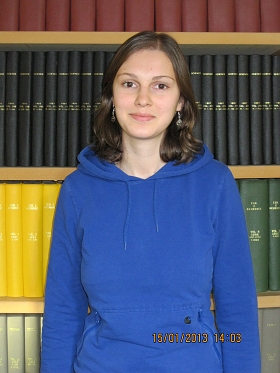
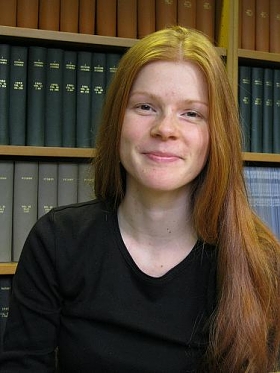
Julia Gottwald has joined Dr Marco Capogna’s group to perform an experimental project in Hilary Term as MSc Neuroscience student. Julia was born in Germany and received a BSc in Biochemistry (with Distinction) at the Free University in Berlin.She has some previous lab experience at the Max Delbruck Center for Molecular Medicine in Berlin, and at the Max Planck Institute for Neurobiology in Munich. Her project aims to investigate the cellular mechanisms underlying the reduction of theta-dominant neuronal oscillations in response to aversive cues in transgenic mice expressing high level of the serotonin transporter (5-HTT OE). She will be studying the cell firing and synaptic transmission, with particular reference to oscillatory activity, in the acute slices of amygdala of wild type and 5-HTT OE mice. This investigation is potentially interesting for fundamental neuroscience, since the serotonin transporter regulates the intensity and duration of serotonergic neurotransmission, but also for preclinical neuroscience, since the serotonin transporter is the principal target for antidepressant and anxiolytic drugs. The electrophysiological experiments in vitro will be performed as part of a collaboration involving Dr. David Bannerman (Exp Psychology), who is testing these mice in freely moving, behaving experiments, and Prof Trevor Sharp (Pharmacology).
/* Style Definitions */
table.MsoNormalTable
{mso-style-name:"Table Normal";
mso-tstyle-rowband-size:0;
mso-tstyle-colband-size:0;
mso-style-noshow:yes;
mso-style-priority:99;
mso-style-qformat:yes;
mso-style-parent:"";
mso-padding-alt:0cm 5.4pt 0cm 5.4pt;
mso-para-margin:0cm;
mso-para-margin-bottom:.0001pt;
mso-pagination:widow-orphan;
font-size:11.0pt;
font-family:"Calibri","sans-serif";
mso-ascii-font-family:Calibri;
mso-ascii-theme-font:minor-latin;
mso-fareast-font-family:"Times New Roman";
mso-fareast-theme-font:minor-fareast;
mso-hansi-font-family:Calibri;
mso-hansi-theme-font:minor-latin;
mso-bidi-font-family:"Times New Roman";
mso-bidi-theme-font:minor-bidi;} - Unit10 Jan 2013
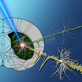
Tommas Ellender, James Harwood, Polina Kosillo, Marco Capogna and Paul Bolam report in the latest issue of the Journal of Physiology (591: pages 257-272, 2013) for the first time the heterogeneous synaptic properties of the two main thalamic projections to the striatum which originate in the rostral central lateral (CL) and the caudal parafascicular (Pf) nucleus. Building on previous work of the Unit showing that the thalamostriatal neurons in these nuclei differ in their morphology, firing properties, as well as their striatal targets (Lacey et al. 2007), we set out to test the hypothesis that synapses formed in the striatum by neurons originating in either nucleus have different functional properties. To address this we isolated and selectively activated the thalamostriatal projections using targeted viral delivery of channelrhodopsin-2 (ChR2). Whole-cell patch-clamp recordings of neurochemically identified medium-spiny neurons (MSN) together with optical activation of specific thalamic inputs enabled us to identify the properties of the two types of thalamic synapse in the adult mouse striatum. We found that thalamostriatal synapses differ significantly in their peak amplitude responses, short-term dynamics, expression of ionotropic glutamate receptor subtypes and potential to exhibit spike timing-dependent plasticity. Our results reveal a hitherto unidentified heterogeneity in the thalamostriatal projection and suggest they subserve different roles in striatal function. More specifically they suggest that CL synapses may act as drivers of MSN spiking activity whereas Pf synapses may subserve the role of modulators of the activity of MSNs.
- Unit7 Jan 2013

We are pleased to welcome Mr Nandor Nemes to the Unit. Nandor has joined Professor Peter Somogyi’s group for a short visit as a gap year trainee from Bristol, and during his time he will learn concepts of brain organisation and participates in the analysis of neuronal circuits in the hippocampus using microscopic methods. He plans to study Neuroscience at Glasgow University.
- Unit19 Dec 2012
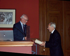
Alan Cowey (life), FRS, FMedSci, Emeritus Professor of Experimental Psychology, Oxford and Honorary Associate of the Unit, passed away on the 19th of December 2012 after being diagnosed with cancer on the 24th of September. Alan was one of Peter Somogyi’s closest friends and past collaborator, a pioneer of visual neuroscience, an unflinching supporter of the Unit, a true gentleman and an example to us all as a scientist and as a person. Alan was elected an Honorary Fellow of the Hungarian Academy of Sciences in 2010 and gave his inaugural lecture at Budapest in April 2011 (see picture). He lectured in the symposium at the Hungarian Embassy in London on the 27th of February 2012: Visions, links and opportunities in Hungarian and British neuroscience.
He is sorely missed.
- Unit14 Dec 2012
d65d.jpg?itok=Ktsbmf2j "Unit members and visitors")
The Unit held its Winter Science Day on Friday 14th December 2012. The event this year was held at the Oxford University Museum of Natural History.
Over 70 Unit members and visitors attended including our colleagues from the Centre for Brain Research in Vienna. There were 12 oral presentations and 28 posters describing on-going projects and plans for the future.
The meeting was followed by drinks in the Museum Court Area and dinner in the Museum Gallery.
The museum was opened in 1860 to house the University Science Departments. It was Oxford’s ‘Cathedral to Science’, and this is certainly reflected in its architecture. On 30th June 1860, the museum hosted the “Great Debate” between Samuel Wilberforce and Thomas Huxley on Darwin’s theory of evolution.
- Unit12 Dec 2012
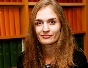
Dr. Stéphanie Trouche joins the Unit as an MRC Investigator Scientist. Stéphanie’s previous research has focused on the neural circuits underlying memory processes and has employed system-neuroscience approach combining behavioural, neuroanatomical and imaging tools. In September 2009, Stéphanie obtained her Ph.D. in Neuroscience at the University of Toulouse, France, where she examined the contribution of new hippocampal neurons to spatial memory processes. She moved in January 2010 as a Fyssen postdoc fellow at Tufts University School of Medicine (Department of Neuroscience, Boston, USA) in the group of Dr. Leon Reijmers where she studied the role of the inhibitory circuits during memory extinction. She will now work in the group of Dr. David Dupret where she will be studying how the assembly firing patterns in the hippocampus contribute to the formation and expression of spatial memory traces.
- Unit3 Dec 2012
We welcome Anne Petzold for an internship in the Unit. Under the supervision of Dr Juan Mena-Segovia, Anne will learn electrophysiological and anatomical techniques, and develop the basis for a project to characterise the interaction between brainstem and midbrain neuromodulatory systems during reward-seeking behaviour.
Anne obtained her Bachelor’s degree in Philosophy of Neuroscience in Germany. Getting initially interested in computational approaches, she soon moved into studying experimental Neurosciences. She concluded her studies with a project on the importance of neurotrophins in hippocampus-mediated spatial learning, graduating with an MSc degree this year. Before coming to the Unit, she worked with Gonzalez de Polaviaja at the Ramón y Cajal Institute in Madrid on a project identifying brain areas relevant for the processing of social information.
- Unit27 Nov 2012
 The cover image shows a light microscopic reconstruction of biocytin-labelled GABAergic neurogliaform cell and glutamatergic principal neuron of the mouse basolateral amygdala complex. Soma and dendrites (thick) of the neurogliaform cell are shown in red, and the axon (thin) is shown in blue; soma and dendrites (thick) and the axon (thin) of the principal cell are shown in black.
 Miroslawa Manko, Thomas Bienvenu, Yannis Dalezios and Marco Capogna report in the last issue of the Journal of Physiology (590.22:5611-5627, 2012) a novel interneuron type of the amygdala, termed neurogliaform cell, and study its function by a combination of in vivo and in vitro techniques. They used a mouse line expressing a green fluorescent protein (GFP) under the neuropeptide Y (NPY) promoter. Paired recordings between presynapticNPY-GFP-expressing (+) cells and postsynaptic principal neurons (PNs) of the basolateral amygdala (BLA) were performed. The NPY-GFP+ neurons displayed small somata and short dendrites embedded in a cloud of highly arborized axon, suggesting a neurogliaform cell (NGFC) type. A NPY-GFP+ cell evoked a GABA-A receptor-mediated slow inhibitory postsynaptic current (IPSC) in a PN and an autaptic IPSC. The slow kinetics of these IPSCs was likely caused by the low concentration and spillover of extracellular GABA. They also report that NGFCs of the BLA fired action potentials phase-locked to hippocampal theta oscillations in anaesthetized rats. When this firing was re-played in NPY+-NGFCs in vitro, it evoked a transient depression of the IPSCs. Presynaptic GABA-B receptors and functional depletion of synaptic vesicles determined this short-term plasticity. Synaptic contacts made by recorded NGFCs showed close appositions, and rarely identifiable classical synaptic structures. Thus, they report a novel interneuron type of the amygdala that generates volume transmission of GABA. The peculiar functional mode of NGFCs makes them unique amongst all GABAergic cell types of the amygdala identified so far.
- Unit22 Nov 2012
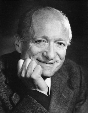 Stephen W. Kuffler, 24th August 1913 – 11 October 1980 (Photograph by: Bachrach Photographers, Boston, Massachusetts, USA)
Peter Somogyi, Unit Director, received the Semmelweis-Budapest Prize 2012 and delivered his lecture “Co-operative chronocircuits in the hippocampus of the brain” at the Hungarian Academy of Sciences at 2.00pm on the 22nd November 2012. He has dedicated the 10,000 Euro prize to the setting up of the Stephen W. Kuffler Scholarship Foundation. Stephen Kuffler was a pioneer genius and an excellent mentor and tutor of talented students, several of whom went on to win Nobel Prizes. He founded the first department of neurobiology in the USA at Harvard University. See highlights of Kuffler's contribution to science in Hubel's Eye, Brain and Vision.
The founding members of the Foundation are:
David H. Hubel, MNAS, Nobel Laureate in Physiology or Medicine (1981)
Eric R., Kandel, MNAS, Nobel Laureate in Physiology or Medicine (2000)
Edward A. Kravitz, MNAS, George Packer Berry Professor of Neurobiology
John G. Nicholls, FRS, Professor of Neuroscience
A. David Smith, FMedSci, Professor Emeritus of Pharmacology
Peter Somogyi, FRS, FMedSci, CMHAS, Professor of Neurobiology
E. Sylvester Vizi, MHAS, Institute of Experimental Medicine, Hungarian Academy of Sciences
Torsten Wiesel, MNAS, 1981 Nobel Prize in Physiology or Medicine
- External9 Nov 2012

Rob Stewart recently completed the 2012 Abingdon Marathon, finishing 64th in a field of 764 runners. Rob achieved a new personal best and unit record time of 2 hours, 57 minutes and 34 seconds.