- Unit3 May 2011
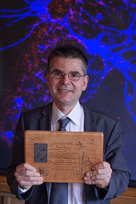
Functional properties of fast spiking GABAergic interneurons in thehippocampal network.Prof. Peter Jonas, Institute of Science and Technology (IST), Vienna, Austria.
Prof. Jonas' Lecture focused on two aspects constantly present in Prof. Smith's scientific thinking and production: exocytosis and GABAergic cells. In his Lecture, the speaker illustrated the mechanisms underlying release of the neurotransmitter GABA by parvalbumin-expressing interneurons of the rat hippocampus. At these synapses, the "nanodomain" coupling between calcium channels and calcium sensors promotes fast and efficient transmitter release. Furthermore, an unexpected small number of calcium channels trigger transmitter release. Finally, GABA released by these interneurons is rapid and highly synchronised. Combined these factors ensure that parvalbumin-expressing GABAergic cells operate with high speed and temporal precision.
The Lecture was set up to celebrate the vision of the previous Chair ofPharmacology Prof. A. David Smith, Honorary Associate Director and founderof the Unit, and the successful conclusion of the latest quinquennialscientific review of the Unit.
To commemorate the Lecture, Prof. Jonas received a laser engraved cherrywood plaque, designed by Unit Artist Ben Micklem and illustrating aspectsof anatomical neuropharmacology at Oxford from molecules to the brain.
- Unit21 Apr 2011
 An excitatory bouton (b) containing multiple VGluT1 immunogold particles makes an asymmetrical synaptic contact (arrowhead) with a dendritic spine.
Published by Jonathan Moss, Mark Ungless and Paul Bolam in the European Journal of Neuroscience.
Abstract: Midbrain dopamine neurons signal rapid information about rewards and reward-related events. It has been suggested that this fastsignal may, in fact, be conveyed by co-released glutamate. Evidence that dopamine neurons co-release glutamate comes largelyfrom studies involving cultured neurons or tissue from young animals. Recently, however, it has been shown that this dualglutamatergic dopaminergic phenotype declines with age, and can be induced by injury, suggesting that it is not a key feature of adultdopamine neurons. Here, we provide further support for this view by showing that dopaminergic axons and terminals in subregions ofthe adult striatum do not express vesicular glutamate transporters (VGluT1, VGluT2 or VGluT3). Striatal tissue from the adult rat wasimmunolabelled to reveal tyrosine hydroxylase (TH; biosynthetic enzyme of dopamine) and one of the three known VGluTs.Importantly, we compared the immunogold labelling for each of the VGluTs associated with TH-positive structures with backgroundlabelling at the electron microscopic level. In addition, we carried out a subregional analysis of the core and shell of the nucleusaccumbens. We found that dopaminergic axons and terminals in the dorsolateral striatum and ventral striatum (nucleus accumbenscore and shell) do not express VGluT1, VGluT2 or VGluT3. We conclude, therefore, that in the normal, adult rat striatum,dopaminergic axons do not co-release glutamate.
- Unit21 Apr 2011
Published by Miroslawa Manko, Raffaella Geracitano & Marco Capogna in the Journal of Physiology.
Abstract: Intercalated cells (ITCs) of the amygdala are clusters of GABAergic cells that surroundthe basolateral complex of the amygdala (BLA). Growing evidence suggests that ITCs are requiredfor the expression of fear extinction. The main intercalated nucleus (Im) is the largest of the ITCclusters and could also be important for emotional processing.We usedwhole-cell recordings fromIm neurons in acute slices of mouse amygdala.We found that these neurons were medium-sizedspiny projection cells. Their passive and active membrane responses were consistent with thosepreviously reported in other ITC clusters. The axon of Im neurons was, in many cases, cut atthe slice boundaries, suggesting long-range projections. Axonal branches could be detected inseveral amygdala nuclei where they made functional synapses. We also functionally studied Imcell inputs. Excitatory postsynaptic currents (eEPSCs) were evoked by the stimulation of theIm, intermediate capsula (IC), external capsula (EC) or BLA, when GABAergic transmission waspharmacologically blocked.An occlusion test indicated that fibres recruited by stimulating ImandIC, or Imand EC were distinct. These eEPSCs had both NMDA and AMPA receptor components.Inhibitory postsynaptic currents (eIPSCs) were evoked after the stimulation of the Im, the ECand the BLA, when glutamatergic transmission was pharmacologically blocked. Furthermore,dopamine reversibly hyperpolarised, and decreased the firing frequency and the input resistanceof Im cells via dopamine type 1 receptor. Our data suggest that the Im is functionally connectedto other amygdala nuclei and is under neuromodulatory influence.We propose that the Imservesas key neuronal substrate of fear extinction.
- Unit21 Apr 2011
Published by Marco Capogna in the Journal of Physiology 589.8: 1875-1883, 2011 (PDF).
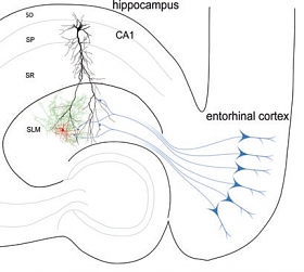
Abstract: The stratum lacunosum moleculare of the hippocampus is an area of integration that receivesinputs from extrinsic excitatory fibres including those from the entorhinal cortex, and is underthe control of several neuromodulators. A critical aspect is the presence in this hippocampal layerof specific interneurons that are likely to influence the strength and the temporal structure ofentorhinalCA1 hippocampal dynamics. I review here recent data on the physiological role ofthese interneurons. Special focus is devoted to one interneuron type, the so-called neurogliaformcell, because recent studies have defined its unusual mode of cell-to-cell communication. Neurogliaformcells mediate feedforward inhibition of CA1 pyramidal cells, form a network of cellsconnected via chemical and electrical synapses, and evoke slow inhibitory synaptic currentsmediated by GABAA and GABAB receptors. The modulation of entorhinal input by neurogliaformcells and their contribution to network theta rhythm are also discussed. I hope thatnovel information on neurogliaform cells will contribute to the ever-growing appreciation ofGABAergic cell type diversity, and will inspire neuroscientists interested not only in synapticphysiology but also in the brain's spatial representation system.
- Unit18 Apr 2011

Megan is joining the Bolam Group as the Girdlers' New Zealand HRC Postdoctoral Fellow. Her research will focus on the analysis of populations of striatal interneurons, initially by electron microscopy.
Megan completed her PhD in Pharmacology at the University of Auckland, New Zealand, under the supervision of Associate Professor Michelle Glass. Her research has centred around the endocannabinoid system in Huntington's disease, in both transgenic mice and post-mortem human tissue, to evaluate the putative pathological nature of changes occurring in this neuromodulatory system.
- Unit11 Apr 2011
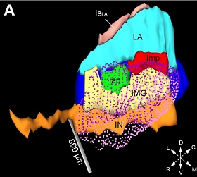
The group of Marco Capogna in collaboration with previous Unit members (Francesco Ferraguti, Yannis Dalezios) publishes on distinct functions of intercalated cell masses of the amygdala in the Journal of Neuroscience, 31 (13): 5131-5144, 2011.
Intercalated cell masses have been implicated in acquisition and consolidation of extinction learning. Busti et al. reports that these neurons are more heterogeneous than previously appreciated. By using cell labelling and electrophysiological techniques, three distinct cell clusters were identified exhibiting distinct axonal projection patterns and differential activation after fear learning and fear extinction.
The paper has been selected and evaluated this week by Gary Aston-Jones, a Member of the Faculty of 1000 (F1000), which places it at the top 2% of published articles in biology and medicine (F1000 evaluation for this paper).
Busti D., Geracitano R., Whittle N., Dalezios Y., Manko M., Kaufmann W., Saetzler K., Singewald N., Capogna M., and Ferraguti F. (2011) Different fear states engage distinct networks within the intercalated cell clusters of the amygdala. Journal of Neuroscience, 31 (13): 5131-5144. (PDF)
- Unit8 Apr 2011

We are pleased to welcome Ms. Maureen Turner to the Unit. Maureen graduated from the University of Arizona with a B.A. in Psychology, and is currently studying for her M.Sc. in Neuroscience at the University of Oxford. Maureen has joined the Magill Group to undertake her second lab rotation, and is using an advanced combination of in vivo electrophysiology and anatomy to elucidate the operational principles of the motor cortex in dopamine-intact and Parkinsonian brains.
- Unit6 Apr 2011

We are delighted to welcome Adam Nusser who has joined Professor Somogyi's group as a visiting student. Adam studies physics at the Budapest University of Technology & Economics.
- Unit14 Mar 2011
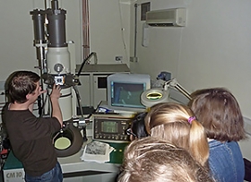
On 14th March the Unit held its seventh annual Open Day for local schools. Four local schools attended the day and 48 sixth form students took part in the activities. After a brief introductory talk by Dr Jeff McIlhinney, which described the breadth of the Unit's work, from molecules to minds, and introduced the students to some basic brain facts, they were able to visit the laboratories. Here they spent time getting hands-on experience with instruments and real specimens, providing a glimpse of what to expect in a working research laboratory. They all visited two of the research groups.
Student feedback indicated that the visit was very stimulating, and that the research they were shown was interesting. Several students asked questions that indicated an interest in how to become a research scientist, as well as what it was like to do research, and how well it was paid. It was clear that the teachers also found the experience stimulating, and we were again told that after last year's visit some students were inspired to make University applications for various bioscience degrees, including neuroscience and medicine. From the students' responses the highlights of the visit included: looking at specimens in the electron microscope, seeing how the brains were sectioned, doing the molecular biology mini-practical, seeing the confocal microscope images, the Parkinson's talk, being told about how to monitor brain activity and how this can represent learning processes. Thanks to all who participated and made this day possible.
- Unit9 Mar 2011

A warm welcome to Judith van Alden, a MSc student in Clinical Neuroscience at University College London carrying out a research project in Prof. Bolam's lab, under the supervision of Dr. Mena-Segovia. Judith will be studying the brainstem neuronal pathways affected in animal models of Parkinson's disease using immunohistochemical and stereological techniques.